Saturday, October 18, 2008

ANTI-SMOKING PILL SHOWS PROMISE IN CURBING DRINKING

Tobacco cessation is the major concern now a days, as "Oral cancer" has created havoc worldwide especially in India.
Many products are available & coming up in the market as a substitute to tobacco.
The latest one is NRT-Nictine replacement therapy in the form of chewing gums, tobacco batches etc. but all of them have limited use & results are nott effective

Friday, October 17, 2008

HANDPIECE MAINTENANCE

For tips on maintaining your handpieces check out this link -

http://rapidshare.com/files/153692751/.pdf_hp_maintanance.pdf_1_.html

FREE MEDICAL BOOKS DOWNLOAD

From the following site:
http://rahulshetty87.googlepages.com/books

Thursday, October 16, 2008

HISTOLOGY OF TOOTH DEVELOPMENT


Bibliography: http://in.youtube.com/watch?v=LVC4_FIEeFs&feature=related

Wednesday, October 15, 2008

TOOTH ERUPTION


Bibliography: http://in.youtube.com/watch?v=0pzIRg8cW4A

CERVICAL CANCER VACCINE ENTERS INDIAN MARKET

Merck Sharp & Dohme (MSD), a wholly owned subsidiary of pharma major Merck & Co. Inc. launched a vaccine to prevent cervical cancer, the most common condition in India with more than 1,30,000 women diagnosed with it every year.
The vaccine is one of its kind & has been approved by the US Food & Drugs Authority.

The vaccine Gardasil was launched in 2006& is available in 108 countries.
Regulatory approvals & Immunogenity studies delayed its launch in India.
Currently, trials are being done by the Indian Council of Medical Research & "could take upto 3 years, but once they are done, the vaccine will be made available by the government as well," said Naveen A. Rao, mabaging director, MSD India.

Gardasil is priced at Rs2,800 ashot with three doses prescribed for efficacy.
According to a World Health Organisation Study, the risk of the cancer in India is 2.4% compared with an average of 1.3% for the world.

Tuesday, October 14, 2008

DENTAL FLUOROSIS

Dental fluorosis is a common disease in Punjab(India).

It is due to an unusually high dose of fluorides during odontogenesis causing a structural modification of hard dental tissues and thereby resulting in a hypomineralisation of these tissues.

Fuorotic enamel is a hypocalcified, porous,brittle and most unaesthetic tissue.


Bleaching has been suggested by several authors in order to treat the unaesthetic aspect of dental fluorosis, but many results are however unsatisfactory.

This is a novel method which is based on the structural characteristics of the fluorotic enamel & organic and exogenous nature of fluorotic enamel stains which includes four different stages:-

1) Cleansing the enamel surface with pumice
2) Enamel etching with hydrochloric acid
3) application of sodium hypochlorite.
4) application of dental adhesives.


INTRODUCTION

A frequent question asked by most of patients residing in the fluorotic belt of Punjab (India) is "will my tooth turn white?"
Usually the answer is a "yes" with the explanation that the modern dental treatment procedures are such as to esthetically synchronise the facial harmony of tooth structure.

The reason for this discoloration is a high fluoride concentration in water in certain areas of Punjab.

The normal colour of permanent teeth is greyish yellow, greyish white or yellowish white but the number of people with this colour are usually limited owing to over aggressive tooth brushing and abrasive cleansing materials, acidic food and drinks and last but not the least,ageing.

The elderly people thus, have more yellowish teeth as compared to younger persons.

These alterations in colour maybe physiologic or pathologic and endogenous or exogenous in nature.

The modern era is an era of esthetics.

People having teeth with normal colour also want to have whiter teeth to improve their smile.

So one cannot ignore the wishes of such patients and hence bleaching, as we know, has emerged as the simplest, most common, least invasive and least expensive means available to dentists to lighten discoloration.


HISTORY

Many agents have been used in the past and a number of new methods have continued to be introduced.

It was oxalic acid first by chappel in 1877 which was followed by various forms of chlorine, until hydrogen peroxide was first used by Harlan in 1884.

Many advances continued focussing basically on the ways to facilitate the absorption of bleaching agent.

The recent developments of hi-tech computer imaging have enhanced patient understanding, expectation and ultimately satisfaction.


MODE OF ACTION


Bleaching works by oxidation in which the bleaching agent enters the enamel &/or dentin of the discolored tooth and reduces the molecules containing discoloration. The bleaching depth depends on the cause of the stains and where and how deep the stain has permeated the tooth structure plus how deep the bleaching agent can permeate to the source of discoloration and remain there long enough to release deep stains.


ETIOLOGY OF TOOTH DISCOLORATION

Extrinsic discolorations are found on outer surface of teeth and are usually Of local origin e.g. Tobacco, paan, tea, coffee,turmeric, silver nitrate stains, oral intake of iron suspensions, continuous use of mouth washes and gum paint.

Intrinsic stains are found within the enamel and dentin and are caused by the deposition of the substances within these structures e.g. Tetracycline, Fluorosis stains, amelogenesis imperfecta, dentinogenesis imperfecta, pulp necrosis etc.


HISTOPATHOLOGY


Fluorosed teeth are also called mottled teeth .

Such teeth appear when child ingests excessive fluoride during enamel formation or calcification in areas where drinking water contains more than 1ppm of Fluoride.

The higher concentration of fluoride is believed to cause a metabolic alteration in the ameloblasts which results in defective matrix & improper calcification.

1 ppm of fluoride has no biological side effects on the vital organs of human body i.e. Kidney, heart & lungs.

Fluoride up to 4ppm in drinking water occasionally produces skeletal fluorosis but above 8ppm coupled with malnutrition positively causes not only skeletal fluorosis but irreversible bone changes & deformity as well.


HISTLOGY


Histological examination shows hypomineralised, porous sub-surface enamel below a well mineralised surface layer.

The most affected teeth (in decreasing order) are premolars, 2ndmolars, followed by maxillary incisors, canines & 1st molars.
Mandibular incisors are affected least.


STAGES OF FLUOROSIS


The appearance of teeth depends upon the severity of the lesion which in turn depends upon the fluoride contents consumed by a particular individual through the water supply.

1) the constant use of water having fluoride to the extent of 1ppm causes mildest grade of mottling in 10% of the population.

2) as concentration of fluoride increases, the effect worsens, so much so that when the concentration reaches 6ppm,incidence of mottling is 100%.

3) very mild :- in this type there are very small white areas occasionally seen on the tooth surfaces, but do not involve more than 25% of tooth surfaces.

(4) mild :- in this type there is more extensive tooth involvement and involves 50% of tooth surfaces.

(5) moderate :- more surfaces are involved here and are subjected to attrition. They show marked wear with yellow or brown pigmentation.

(6) severe :- all enamel surfaces are involved, so much so that the tooth morphology is affected.there is discrete or confluent pitting of enamel surfaces.
Brown stains are widespread & the tooth often presents a corroded surface.


OPTIMUM FLUORIDE LEVELS:

In cold climate, recommended fluoride levels may be as high as 1.2 ppm whereas in extremely hot climate, a level of 0.7 ppm is recommended.
In moderate climate, the optimum fluoride level is 1 ppm.

High temperature causes increase in mottling because there is increased consumption of water containing fluoride.

Distribution of mottling in various areas of teeth has no relation with periods of mineralisation of crown.

Teeth are only affected provided the child lives in the area of fluorosis during the time of enamel mineralisation.

Brown tooth stains respond to treatment but white stains are not effectively resolved.

It has been observed that teeth in process of eruption receive maximum benefit from optimum amount of fluoride plus teeth exposed to fluoride shortly after eruption were also protected although to a lesser degree.


DIFFERENT FLUORIDE LEVELS IN PUNJAB & OTHER STATES OF INDIA


I) Punjab
1) bhatinda - 4.5 ppm
2) mansa - 4.2 ppm
3) mukatsar - 3.3 ppm
4) faridkot - 3 ppm
5) ferozepur - 2.6 ppm
6) moga - 2 ppm
7) sangrur - 1.35 ppm
8) jalandhar - 0.55 ppm
9) amritsar - 0.45 ppm 10) hoshiarpur - 0.44 ppm
11) nawanshahar - 0.4 ppm
12) fatehgarh sahib - 0.37 ppm
13) patiala - 0.35 ppm
14) ropar - 0.3 ppm
15) kapurthala - 0.25 ppm
16) ludhiana - 0.22 ppm
17) gurdaspur - 0.15 ppm

II) Andhra Pradesh
1) nalgonda - 20.6ppm
2)prakasan - 12.0ppm
3)vishakhapatnam - 11.0ppm
4) anantpur - 10.1ppm
5)guntar - 10 ppm
6)medak - 9.8ppm
7)kunoor - 9.6ppm
8)nellore - 8 ppm
9)mehboobnagar - 6.4 ppm 10)warrangal - 5.8 ppm
11) kareemnagar - 4.9 ppm
12) hyderabad - 4.8ppm
13) cuddapah - 4.6ppm
14) nizamabad - 3.0ppm
15) chittoor - 2.9 ppm
16) adkabab - 2.8 ppm
17) srikakalam - 2.8 ppm
18) Godavari - 1.6 ppm

III) Gujarat
1) kutch - 1.2 -- 11 ppm
2) bhavnagar - 1.5 - 4ppm
3) jamnagar - 1.5 - 4ppm
4) rajkot - 2.5ppm
5) saurashtra - 1.5 - 2.5 ppm
6) rajpur - 0 - 2.5 ppm 7) banakanta - 1.5 - 2ppm
8) godar - 1.6 - 1.7ppm
9) godhra - 0 - 1.6 ppm
10) surinderanagar - 0 - 1.5 ppm
11) surat - 0 - 1.3 ppm

IV) Tamil Nadu
1) Coimbatore 2) Dharampur 3) Madurai 4) Narkot 5) Salem 6) Trichi
all 1.5 --- 5ppm

V) Kerala
1) Allepey 2) Eranakulam 3) Quillon 4) Trichur
all 0 -- 1.5 ppm

V) Rajasthan
1) Bharatpur - 28ppm
2) Tonk - 21ppm
3) Alwar - 20.6ppm
4) Sikar - 19.1ppm
5) Ajmer - 18.4ppm
6) Bhilwara - 16.5ppm
7) Swaimadhopur - 16.1ppm
8) Jhalawar - 16ppm
9) Churu - 16ppm
10) Jodhpur -16ppm
11) Sirohi - 15.8ppm
12) Jaipur - 15ppm
13) Nalpur - 14.2ppm
14) kota - 14.2ppm
15) dungarpur - 12ppm
16) bikaner - 10.2ppm
17) barmer - 10ppm
18) pali -- 9.1ppm
19) ganganagar - 9ppm
20) jalour -- 8ppm
21) wagpur - 7.1ppm
22) chittorgarh - 6ppm
23) bundi - 5.8ppm
24) banswara - 4.3ppm
25) jhunjhunu - 2.2ppm

VI) Uttar Pradesh
1) Gorakhpur - 0.6-6.8ppm
2) Shahjahanpur - 4ppm
3) Lakhpur -0.1-4ppm
4) Rai bareilly - 0.6-3ppm
5) Banda - 0.6-3ppm
6) Agra - 0.2-3ppm
7) Kanpur - 0.2-3ppm
8) Varanasi - 0.2-3ppm
9) Unna - 0.1-3ppm
10) Aligarh - 0.4-2ppm
11) Allahabad - 0.2-2ppm
12) Itah -0.8-1.6ppm
13) Hamirpur - 0.6-1.6ppm
14) Azamgarh - 0.1-1.6ppm
15) Muradpur - 1.0-1.4ppm
16) Jamalpur - 1.0-1.2ppm
17) Lucknow - 0.8-1.2ppm
18) Meerut - 0.4-1.2ppm
19) Bulandshahar - 0.4-1.2ppm
20) Dijnor - 0.2-1.2ppm
21) Jhansi - 0.2-1.2ppm
22) Bareilly 0.1-0.9ppm
23) Balliya - 0.4-0.8ppm
24) Barabanki - 0.4-0.8ppm 25) fatehgarh - 0.4-0.8ppm
26) mirzapur - 0.4-0.8ppm
27) gadhepur - 0.3-0.8ppm
28) gonda - 0.2-0.8ppm
29) basti - 0.2-0.8ppm
30) jalpum - 0.1-0.8ppm
31) dehradun - 0.1-0.8ppm
32) pratapgarh - 0.4-0.6ppm
33) manipuri - 0.4-0.6ppm
34) lahtpur - 0.1-0.6ppm
35) muzaffarnagar - 0.2-0.5ppm
36) rampur - 0.2-0.4ppm
37) pilibhit - 0.2-0.4ppm
38) bijnor - 0.1-0.4ppm
39) fatehabad - 0.1-0.4ppm
40) badari - 0.1-0.4 ppm
41) sitapur - 0.1-0.4ppm
42) saharanpur - 0.1-0.4ppm
43) mathura - 0.1-0.4ppm
44) faizabad - 0.2ppm
45) etawah - 0.1-0.2ppm
46) nainital - 0.1-0.2ppm
47) dahrich - 0.1-0.2ppm
48) sultanpur - 0.1ppm


TREATMENT OPTIONS

Basically for all these stains or in particular fluorotic stains the treatment options available to us include :-

1) Veneering / laminates or placement of porcelain crowns
2) Micro / macroabrasion
3) Bleaching -
a) vital tooth inoffice bleaching
b) nightguard home bleaching
c) our novel method of inoffice bleaching


1) VENEERING OR LAMINATES OR CRAMIC CROWNS

Advantages:

1) esthetically more acceptable
2) Long lasting
3) Durable
4) Simple
5) Can be given over endodontically treated tooth
6) more strength and resistance to forces

Disadvantages:

1) brittle
2) less shear strengh
3) causes loss of tooth structure
4) patient may not be willing
5) susceptible to fracture
6) due to tooth reduction, pulp & other tissues may face trauma
7) overcontouring may make it appear & feel unnatural
8) vitality tests cannot be done once crowns are properly fit
9) post cementation caries difficult to detect
10) lab.procedure needs precision for proper marginal seal
11) gingival irritation- may cause hyperaemia & bleeding


2) MICRO / MACRO ABRASION:-

This technique involves applying of 18% hcl to soften the enamel And then abrading it with a controlled abrasive technique With pumice to remove superficial stains / defects. Instead of pumice, even silicon carbide may be used with 11%hcl.

Advantages:

1) improved method for superficial stains
2) safer method
3) involves physical removal of tooth structure

Disadvantages:

1) can cause sensitivity
2) causes wearing of tooth structure
3) patients might not allow cutting of tooth structure
4) defect may persist after finishing of technique for which a restorative alternative is needed


3) BLEACHING:-

This procedure has many methods and techniques involving various solutions in each technique.

Advantages:


1) easy
2) time saving
3) cheaper
4) patient acceptance better
5) can be carried out both in office & at home

Disadvantages:


1) requires patient cooperation(especially for home bleaching)
2) cannot be used where teeth have large pulps
3) cannot be used where teeth are too dark
4) cannot be used where the patient expectations are too high
5) cannot be used in impatient patients
6) causes cervical resorption
7) cannot be used in attritioned teeth which might cause sensitivity
8) cannot be used where teeth are bonded, laminated or have extensive restorations
9) not a perfect technique & merely changes colour to variable depths
10) lasts for only 1 - 3 years (short period)


A) Vital tooth inoffice power bleaching

This technique uses a combination of 37% phosphoric acid & 35%hydrogen peroxide.the oxidation reaction is generally promoted by a heated instument or with intensive light.in this method, one application is carried out weekly for 2 - 6 appointments with each treatment lasting 30 minutes. Use of phosphoric acid by this technique is optional.

Advantages:

1) caustic chemicals are totally under dentist's control.
2) soft tissue protection is better achieved by dentist.
3) bleaching of tooth is achieved more rapidly

Disadvantages:

1) slightly costly procedure.
2) unpredictable results.
3) uncertain duration of treatment
4) soft tissue damage possible for both dentist & patient.
5) rubber dam causes discomfort.
6) can cause post operative sensitivity.


B) Night guard home bleaching

This procedure involves making an impression of the teeth & pouring a cast of the same, trimming of the cast, application of a blockout resin & fabrication of a night guard tray by a vaccum former machine.
After cooling, the tray is trimmed & a 10 - 15% gel of carbamide peroxide is recommended for the same.
In this procedure the total treatment time is 2 - 6 weeks.

Advantages:

1) use of lower concentration.
2) ease of application.
3) minimal side effects.
4) lower cost (as compared to veneers)
5) lesser chair time.
6) much lesser labour intensive.

Disadvantages:

1) have to rely a lot on patient compliance for results.
2) longer treatment time.
3) unknown potential for soft tissue changes with excessive use.
4) treatment results are time & dose dependent.
5) peroxide solution may cause irritation of gingival papilla.
6) teeth become sensitive to temperature changes.


Another method using macken's solution has been described
1 part anaesthetic ether 0.2 ml - removes surface debris 5 parts hcl 38% 1ml --- etches 5 parts hydrogen peroxide 30% 1 ml --- bleaches


Our Approach For Inoffice Bleaching

Indications:

1) Fluorosis stains / systemic fluorosis
2) Tetracycline stains

Contra indications:

1) Hyperaemic gingiva
2) Persistant periodontal problem cases
3) Fractured incisors / anteriors


CLINICAL APPLICATION


The various steps are
1) Cleansing
2) Isolating
3) Etching
4) Rinsing
5) Dehydration
6) Application of solution
7) Scrapping
8) Rinsing
9) Filling


The Steps in detail:
1) cleansing the tooth surface with a nylon tooth brush & a mixture of pumice and water to remove surface debris.

2) isolation is done by application of rubber dam.

3) then dry the tooth surface & do enamel etching with 35% hcl for 20 - 25 seconds.

4) copious rinsing is done to eliminate acid residues & the tooth is subjected to thorogh drying.

5) application of 95% ethyl alcohol to dehydrate the enamel surface.

6) now,the application of 30% hydrogen peroxide(h2o2) is done first for 1 minute followed by alternative application of 5.25% sodium hypochlorite (naohcl) is done for 5 minutes during which it can be re-applied to the tooth surface to keep it wet.

7) the removal of staining molecules can be accelerated by gently scrapping the tooth surface.

8) this is followed by thorough rinsing of tooth surface.

9) this procedure is repeated at the interval of three days for successive sittings till the results are satisfactory.

10) in the end, fill the microcavities caused in the tooth by this solution with a light cure dental adhesive.


Advantages:

1) HCl etches enamel,but does not penetrate.
2) Tooth structure is not damaged.
3) Very very few chances of post - operative sensitivity of tooth.
4) No heat / application is required.
5) Very economical as all the three solutions in quantity of 50 ml. Each cost rs. 250 - 300 (total ).
6) Very low quantity of solutions required at each sitting.


Disadvantages :

1) Fluorosed teeth require larger & repeated sessions to decolorise Them.
2) Some blanching of gingiva can occur which is reversible within Half an hour.
3) Transitory decrease in bond strengh occurs when composite is applied to bleached / etched enamel.however,after a week,no decrease is seen.
4) Unknown duration of treatment.


DISCUSSION

The different hypothesis concerning the fluorotic stains removal are:

1) if a fluorotic tooth is put into a NaOHCl solution,it removes all the stains within a few hours.this confirms the organic & exogenous nature of fluorotic tooth stains which are due to elementary impregnation of a hypocalcified & porous tissue. said by :- Triller m. Alterations des tissues by marie curie in 1984.

2) scanning electron microscope study (sem) study shows that Posteruptive calcified layer covers the fluorotic enamel surface ; hence the mineral layer removal is essential.


CONCLUSION

In the end, i would like to conclude that this system of stains removal seems to be clinically applicable & satisfactory with minimal abrasion of enamel surface to make this technique Universally acceptable , lot of cases have to be treated with this technique.

TWO MINUTE BRUSHING HELPS ACHIEVE CLEANER TEETH: STUDY

NEW YORK (Reuters Health) - Although hard work tends to pay off in other areas of life, forceful toothbrushing appears to be no better at ridding the mouth of plaque than a medium effort.

A group of European researchers discovered that the most efficient means of reducing plaque appears to be brushing for about two minutes at a medium force.

More vigorous teeth cleaning may actually do more harm than good, said Dr. Peter A. Heasman of the University of Newcastle upon Tyne, UK. Research suggests that heavy brushing can damage gums and wear down teeth, both potentially serious oral health problems, he said.

"Although we found that you have to brush your teeth reasonably long and hard to get rid of the harmful plaque which causes dental diseases, our research shows that once you go beyond a certain point you aren't being any more effective," Heasman said in a statement.

"You could actually be harming your gums and possibly your teeth," he added.

Heasman and his colleagues designed the study, published in the Journal of Clinical Periodontology, to determine the most efficient way to brush away plaque. Plaque is a sticky substance that can contain more than 300 species of bacteria, which adhere to tooth surfaces and produce cavity-causing acid. Plaque is a leading cause of gum disease.

During the study, Heasman and his colleagues measured plaque levels in the mouths of 12 people after they brushed their teeth using four different forces and for four periods of time -- 30 seconds, 60 seconds, 120 seconds, and 180 seconds.

The study participants brushed using a power toothbrush, which exerted set forces of between 75 grams and 300 grams. All spent 24 hours without cleaning their teeth before testing how well each technique stripped their mouths of plaque.

Heasman said that a force of 75 grams feels much lighter than one of 300 grams. However, he recommended that people visit their dentist to determine how different brushing forces feel.

"It is very difficult for a lay person to differentiate between brushing forces," Heasman told Reuters Health.

Longer brushing generally appeared better, but the researchers found that 120 seconds of brushing was roughly just as effective at removing plaque as longer brushing. And during those longer sessions, people removed about the same amount of plaque using a force of 150 grams as when they employed forces of 225 and 300 grams.

Although different people may require more or less time to get at all the plaque-ridden nooks and crannies in their mouth, spending around two minutes brushing your teeth seems "about right", Heasman said.

And applying a force beyond 150 grams -- somewhere in between light and forceful brushing -- "offered little benefit to plaque removal," Heasman added.

Furthermore, in toothbrushing, it is possible to have too much of a good thing, the researcher said.

"In the short term, gum changes may become apparent, but in the longer term, tooth wear or toothbrush abrasion is likely with too abrasive a technique, toothpaste, brush or force," Heasman said.

SOURCE: Journal of Clinical Periodontology 2003;30:409-413.

BRUSHING RIGHT AFTER DRINKING SODA (CARBONATED DRINKS) MAY HARM THE TEETH

BERLIN (Reuters Health) - If you rush to brush your teeth right after drinking soda (aerated cold drinks), think again.
Doing so may actually do more harm than good, and it's better to wait 30 or 60 minutes before brushing, according to new research.


Because carbonated drinks are highly acidic and have the potential to damage a tooth's enamel, dentists at Goettingen University, Germany, conducted a study to determine the best time to brush after drinking such beverages.
They found that later -- rather than immediate -- brushing is between three and five times more effective at protecting enamel from the erosive effects of carbonated drinks.


In the study, 11 volunteers wore a sterilized piece of tooth-like material in a removable prosthesis for three weeks.
This was removed in the mornings and evenings and soaked for 90 seconds in a liquid similar in acidity to soda.


Afterwards, the prosthesis was brushed using an electric toothbrush at different times after the 'drink.'
Three weeks later, the researchers measured the thickness of the enamel to see how much damage had been inflicted on the 'tooth.'


Professor Thomas Attin, director of the university's department for tooth protection, preventative dentistry and periodontology, said, "The loss of material was less when the participants waited with cleaning for between 30 and 60 minutes."


Professor Attin presented the research at the annual meeting of the German Association for Tooth Protection, where it was awarded a prize from chewing gum firm Wrigley.


He said tooth enamel appears to suffer less damage when brushing occurs after the tooth has had time to mount its own defence against acidic erosion.


Acidic substances attack tooth enamel, he said, and upper layers of the tooth can even be dissolved in some acidic drinks. However, protective agents in saliva may help repair and rebuild damaged tooth enamel.

Waiting for a while seems to give the teeth a chance to rebuild, the researchers said, while immediate cleaning of such teeth can increase the damage by literally brushing off the affected layers.

CHLORHEXIDINE MOUTHWASH

Chlorhexidine has been proven to be the most effective chemical agent for the reduction of plaque and gingivitis.

1. Reduces pellicle formation.

2. Alters bacterial cell wall causing lysis.

3. has High degree of substantivity i.e., it adheres to tissues and remain for a long time, increasing its effectiveness in fighting bacteria.

5. Non specific antimicrobial activity of chlorhexidine has not been associated with development of resistance of pathological oral bacteria.

6. Due to its fungicidal action, chlorhexidine may benefit HIV infected people because of their propensity to contract oral candidiasis.


As a mouth rinse to control plaque and gingivitis, it is recommended to be used twice daily.
It should not be used within 30 to 60 minutes of a tooth paste since most tooth pastes contain sodium laural sulphate, which can deactivate chlorhexidine.
Also, stannous fluoride products should always be used after chlorhexidine since stannous ion and chlorhexidine both compete for and occupy the same site on the tooth.

Some side effects of chlorhexidine include staining of teeth reversible desquamation in young children, alteration of taste.

Sunday, October 12, 2008

DRY SOCKET

Dry socket is a condition which affects millions of patients around the world specially those who undergo tooth extraction.
This painful event can be avoided in majority of cases by proper understanding. It will then save unnecessary agony to patients and loss of countless hours of dentist's practice in dealing with it.


This condition occurs after tooth extraction, particularly after traumatic extraction, resulting in a dry appearance of the exposed bone in the socket, due to disintegration or loss of the blood clot.
It is basically a focal osteomyelitis without suppuration and is accompanied by severe pain (alveolalgia) and foul odor.
It is also called alveolar osteitis and alveolitis sicca dolorosa. (Dorland, 27th Ed)


The most common, most dreaded and most painful complication of tooth extraction. Clinicians call it a "dry socket" a misnomer that fails to stress the importance of infection in its etiology.
Affected person complains of unbearable pain and sensitivity of intake of food or drinks.


Dry socket usually develops after 3 to 5 post surgical days.
The pathogenesis of dry socket (also called fibrinolytic alveolitis) is a subject of debate with two main opinions.
The first one is based on the presumption that there is a absolute absence of blood clot.
According to the second opinion there is initial blood clot formation, which subsequently gets lysed leaving behind an empty socket.
Streptococci have been implicated as causative organisms, but lysis might occur without bacterial presence also

The following factors are considered important in causation of dry socket:

1. Insufficient blood supply to the alveolus.

2. Preexisting infection. (Granuloma, periodontal or pericoronal infection)

3. Use of large amounts of local Anesthetic, leading to vasoconstriction.

4. Post operative bleeding.

5. Trauma to alveolus during extraction.

6. Infection during or after extraction.

7. Root/bone fragments or foreign bodies left in the socket.

8. Excessive irrigation and curettage.

9. Fibrolytic or proteolytic activity in the clot.

10. Loss of clot due to patient's negligence

11. Predisposing factors in patient, eg smoking, poor general health

12. Dry socket is more often seen in the mandibular molars particularly the third molars. This condition is associated with excruciating pain, foul breath, unpleasant taste, empty socket and gingival inflammation and Lymphadinopathy.


By avoiding all possible averse factors, risk of dry socket becomes less.

Prophylactic packing of alveolus with medicated dressing and advising patient to use 0.2% chlorhexidine mouth rinse may be helpful to avoid dry socket in suspect cases.


Treatment of dry socket is mainly done to control pain by analgesics, advice warm saline rinse to remove food debris, dressing the cavity to protect & heal the socket.
In early stages just initiating fresh bleeding in the socket and giving a pack will resolve this condition.


Zinc oxide dressing also have been advised.


With these precautions and treatment the pain should reduce and granulation of the socket should be observed.
Antibiotic therapy may be used if desired.
Most sockets resolve in 4-5 days.


USEFUL Tips:

1. Always compress the socket after extraction so that chances of clot retention are better.

2. Give all instructions to patient so that he does not disturb the extraction site wound.

3. Ask smoker to stop until extraction wound has healed.

Body has great healing capacity, avoid unnecessary routine irrigation of socket with antiseptic solutions.

If dry socket develops do a simple trick it works for 99.99% cases, take a sharp sickle scaler and scrap the gums surrounding socket, let fresh blood fill the socket, give a pack (wet squeezed gauze)
FAMDENT SHOW 2008
13-16 NOVEMBER, 2008,
WORLD TRADE CENTRE, MUMBAI

AND AVAIL OF THE CASH BACK OFFER (CHECK ATTACHED BROCHURE FOR DETAILS) & MUCH MORE!

REGISTRATION FEE & OFFER INCLUDES:

DELEGATE / PG STUDENT FEE:

Rs. 1100/- (includes Trade Fair, Scientific Sessions, Tea/ Coffee)

Rs. 1900/- (includes Trade Fair, Scientific Sessions, 2 Lunches, Tea/Coffee, Gift, 3 VCD's - one each on Endodontics, Implants & Cosmetic Dentistry)

UNDERGRADUATE STUDENT FEE:

Rs. 850/- (Includes Trade Fair, Scientific Sessions, Tea/Coffee)

Rs. 1650/- (includes Trade Fair, Scientific Sessions, 2 Lunches, Tea/Coffee, Gift).

SO HURRY & SEND A CONFIRMATORY EMAIL TO famdentresponse@gmail.com BY 10TH OCTOBER, 08 TO AVAIL OF THE OFFER. REGISTRATION FEE CAN BE SEND BY 20TH OCTOBER, 08

JOIN THE HUNDREDS OF DELEGATES FOR THE MOST HAPPENING DENTAL EVENT OF 2008!!

For more details on Speakers & Hands-On check the attachment of Famdent Show 2008, fill the attached form with brochure & send us along with Cheque / DD on the address given below.

OR

Payment can also be made at any branch of ICICI Bank into

Account Name: Famdent

A/C no: 026305000519

ICICI Bank Lokhandwala Branch, Mumbai

On depositing please inform us by email and also send us a copy of the payment slip along with the completed Registration form.

FAMDENT,
7/102, Sapphire Court, Azad Nagar
Behind Apna Bazar, J.P. Road,
Andheri (W), Mumbai 400053, INDIA.

Tel: +91 22 65049697 / 22 26732260
Telefax: +91 22 26742425
Email: famdentresponse@gmail.com
Website: www.famdent.com

Saturday, October 4, 2008

TONGUE SCRAPING


Tongue scraping is a daily health-maintenance procedure that very few people bother with, probably because they don't yet realize they need to keep their tongues clean.
However, maintaining fresh breath & healthy gum tissue means you have to remove plaque on a regular basis to keep both your breath & tongue fresh & healthy; food debris, odorous dead bacteria & skin cells, as well as stagnant oral fluids have health & social consequences that are largely avoidable.
Nobody wants bad breath or gum disease, & this section will show you how to take better control of both problems through the use of a tongue scraper.

Why should we bother to scrape our tongues?
First, it is important to realize that the tongue doesn't have a smooth surface, as most people assume.
Rather, it is covered by wormlike projections called filiform papillae that can be long or short, & generally number in the thousands.
One way to visualize these filiform papillae is to compare them to a shag rug.
The wet, not-very-clean shag is what the upper surface of the tongue is like in the majority of people.
The larger surface area of the tongue may harbor many more micro-organisms; there can be literally trillions of living, dying, rotting, stinking micro=organisms on top of the tongue.
Dead skin cells, food debris, & oral fluids are literally saturating the area around the papillae, intermingling with the innumerable micro-organisms -- & they are most definitely not harmless or unnoticed by others (for e.g. bad breath)
Whether the stew is on the tongue or trapped in the sulci, it still stinks, still causes soft tissue breakdown, & still creates inflammation, sometimes dramatically.
Sometimes patients' tongues will be quite tender, & will bleed on tongue scraping efforts-not surprising considering that the same tissue-destroying enzymes & odors that are under the gums in the sulci are in the stew on the top of the tongue.
The tenderness & bleeding usually abate quickly once the tongue gets cleaned daily.

Most scrapers come either as a plastic strip with serrated edges, or as a single-handled metal or plastic device.
Tongue can be cleaned with the toothbrush also.

When scraping the tongue, it is very important to scrape as far back as possible, since getting rid of micro-organisms & odorous debris depends on how effectively this step is performed.
The gag reflex can be a problem for some just starting this important habit, but it is usually overcome quickly & generally ceases to be a problem.
Having a healthy mouth & fresh breath can be a powerful motivator.

How do you know when your tongue is clean?
You'll know when you are scraping & no longer getting any odorous residue, & when your tongue is totally pink


fig. 1. surface of tongue with VSCs & gram negative bacteria
fig. 2. toothbrushing just breaks apart bacteria
fig. 3. scraper sweeps away gram negative bacteria
fig. 4. Rinse clean the deep fissures

Friday, October 3, 2008

DOWN'S SYNDROME / MONGOLIANISM / MONGOLISM / TRISOMY 21


Down syndrome (DS), also called Trisomy 21, is a condition in which extra genetic material causes delays in the way a child develops, both mentally and physically.
It affects about 1 in every 800 babies.

The physical features and medical problems associated with Down syndrome can vary widely from child to child.
While some kids with DS need a lot of medical attention, others lead healthy lives.

Though Down syndrome can't be prevented, it can be detected before a child is born. The health problems that can go along with DS can be treated, and there are many resources within communities to help kids and their families who are living with the condition.


Normally, at the time of conception a baby inherits genetic information from its parents in the form of 46 chromosomes: 23 from the mother and 23 from the father.
In most cases of Down syndrome, a child gets an extra chromosome 21 — for a total of 47 chromosomes instead of 46.
It's this extra genetic material that causes the physical features and developmental delays associated with DS.

Although no one knows for sure why DS occurs and there's no way to prevent the chromosomal error that causes it, scientists do know that women age 35 and older have a significantly higher risk of having a child with the condition.
At age 30, for example, a woman has about a 1 in 900 chance of conceiving a child with DS.
Those odds increase to about 1 in 350 by age 35.
By 40 the risk rises to about 1 in 100.


Kids with Down syndrome tend to share certain physical features such as a flat facial profile, an upward slant to the eyes, small ears, and a large or protruding tongue.


Low muscle tone (called hypotonia) is also characteristic of children with DS, and babies in particular may seem especially "floppy." Though this can and often does improve over time, most children with DS typically reach developmental milestones — like sitting up, crawling, and walking — later than other kids.


At birth, kids with DS are usually of average size, but they tend to grow at a slower rate and remain smaller than their peers.
For infants, low muscle tone may contribute to sucking and feeding problems, as well as constipation and other digestive issues.
Toddlers and older kids may have delays in speech and self-care skills like feeding, dressing, and toilet teaching.


Down syndrome affects kids' ability to learn in different ways, but most have mild to moderate intellectual impairment.
Kids with DS can and do learn, and are capable of developing skills throughout their lives.
They simply reach goals at a different pace — which is why it's important not to compare a child with DS against typically developing siblings or even other children with the condition.

Kids with DS have a wide range of abilities, and there's no way to tell at birth what they will be capable of as they grow up.


Dental association with DS
Introduction:
The orofacial and skeletal development associated with Down's Syndrome contribute to dental problems. It is important to be aware of the type of anatomical soft tissue and dental anomalies which are part of the typical developmental pattern of people with Down's Syndrome, which have influence on dental problems.

Anatomical development changes the cranial base, the mid third of the face and the proportion between the maxilla and mandible. This alteration of the skeleton leads to people with Down's Syndrome having a recognisable facial appearance. The soft tissue feature most affected is the tongue, which is fissured and protrusive. The tongue appears large because it has to rest in a narrow dental arch. The tonsils and adenoids are also enlarged.

Dental anomalies are related to the tooth morphology in that there is:

* Decreased root to crown ratio
* Decreased tooth size
* Hypodontia or partial anodontia
* Delayed eruption


DENTAL PROBLEMS:


The normal development of oral structure and function is altered leading to compromised development of suckling, swallowing, mastication and speech; and to drooling unless there is effective intervention.

The degree of difficulty varies from person to person:


Preventive measures and therapy are needed to ameliorate the problems found in swallowing and mastication. Here an integrated approach can be adopted with the Speech and Language Therapist.


DENTAL DISEASE


People with Down's Syndrome are prone to the same degree of dental disease as the general population.

Periodontal disease
: People with Down's Syndrome develop more severe forms of periodontal disease than the general population.
This may be related to immunological deficiency factors.
This disease is most rampant in young people between 16 and 20 years old.
The progression of the disease gives rise to periods of acute infection and pain, which may result in changes in behavior, refusal to eat or swallowing food whole.

Caries: Various studies have shown a reduced incidence of caries in children and young adults with Down's Syndrome.
This may be due to the fact that many of these children are under supervision in regard to their diet in order to prevent their tendency to obesity.
This is where the dentist and the dietician can work together to make sure the food being consumed is working towards oral and general health improvement.

Risk associated with infection is raised in people with Down's Syndrome as the incidence of congenital cardiac disease is increased in this group (3% to 40%), resulting in a serious risk of endocarditis.

The gag reflex can occur even in the anterior portion of the oral cavity. Any further back than the premolars a gag reflex may be accompanied by a gastro-oesophegal reflux. Children find this most uncomfortable.

Bruxism occurs in people with Down's Syndrome and may be triggered by a state of chronic anxiety, dental malocclusion, temporo mandibular joint dysfunction due to laxity of the supporting ligaments, and/or underdeveloped nervous control.

Dental trauma is frequently experienced due to lack of motor development.
Fracture or luxation of the anterior teeth is frequent and often involves loss of tooth vitality.

TREATMENT & PREVENTION

* Good oral hygiene and supervised tooth brushing programmes
* Education, e.g. via videotapes
* Diet, communication and use of oral muscles. This requires an integrated approach to care, as it involves a team of professionals and carers.
* Management of any malocclusion requires a multi-disciplinary team to carry out diagnosis and treatment planning (e.g. Orthodontist, Restorative and Oral Maxillo-Facial Surgeons)

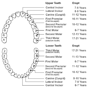
ERUPTION CHART FOR THE PERMANENT TEETH


(CHECK THE CHART FOR ERUPTION OF MILK TEETH UNDER THE POST "MILK/DECIDUOUS/CHILDREN'S TEETH)

Thursday, October 2, 2008

INTERCEPTIVE ORTHODONTICS & SPACE MAINTAINERS

Guidance of the eruption and development of the primary and permanent dentitions is an integral part of the care of pediatric patients.
Such guidance should contribute to the development of a permanent dentition that is in a harmonious, functional and esthetically acceptable occlusion.

Diagnosis and Treatment Planning


Dentists have the responsibility to recognize, differentiate, and either appropriately manage or refer abnormalities in the developing dentition as dictated by the complexity of the problem and the individual clinician’s training, knowledge, and experience. Early diagnosis and successful treatment of developing malocclusion can have both short-term and long-term benefits while achieving the goals of occlusal harmony, function and dental facial aesthetics.

The variables associated with the treatment of the developing dentition which will affect the degree to which treatment is successful include, but are not limited to:

* chronological, mental and emotional age of the patient, to the extend that this affects the ability of the patient to understand and cooperate in the treatment
* intensity, frequency and duration of an oral habit
* parental support for the treatment
* compliance with clinician's instructions
* craniofacial configuration
* variations in craniofacial growth
* concomitant systemic disease or condition
* accuracy of diagnosis
* appropriateness of treatment

Many unpredictable factors can affect the management of the developing dental arches and minimize the overall success of any treatment. These factors cannot always be controlled by the dentist. Appropriate pretreatment records should include those deemed necessary by the individual clinician to adequately diagnose the patient’s condition.

Clinical examination should include:

* an assessment of overall oral health
* facial analysis to determine the growth pattern present
* functional analysis to determine the presence of any deleterious habits and or occlusal dysfunction


Diagnostic records (depending upon the clinical situation) may include:

# extraoral and intraoral photographs to establish a data base for documenting changes during treatment
# diagnostic dental casts to assess the occlusal relationship, determine the arch length requirements for intra and inter-arch tooth size relationships
# intraoral and panoramic radiographs to establish dental age, assess eruption problems, estimate size and presence of unerupted teeth and identify dental anomalies and/or pathology
# cephalometric analysis to determine dental and skeletal relationships and establish a baseline growth record


Space Maintenance

Whenever primary or permanent teeth are lost prematurely and arch integrity is lost, loss of space and arch length, perimeter, or circumference may result. Migration of primary and/or permanent teeth can occur and the available space may be reduced by an amount sufficient to cause some degree of crowding in the permanent dentition.


Indications:

* The premature loss of primary molars may require the placement of a space maintainer to prevent the migration of the adjacent teeth, depending upon the teeth present and the arch length.
* When loss of a primary canine occurs, the dental arch midline may be compromised and the arch length also may be reduced. The premature loss of primary canines may therefore require the placement of a space maintaining appliance to prevent midline deviation and/or loss of arch length, perimeter and/or circumference.
* The premature loss of primary incisors does not usually require the placement of a dental appliance for the maintenance of space because mesial movement of the adjacent teeth is not generally expected.

Contraindications:


* A space maintainer is usually not necessary if there is a sufficient amount of space present to allow for eruption of permanent tooth/teeth.
* A space maintainer may not be recommended if severe crowding exists, such that space maintenance is of minimal effect and subsequent orthodontic intervention is indicated.
* A space maintainer may not be necessary if the succedaneous tooth will be erupting soon.


Types of Space Maintainers


The treatment modalities may include, but are not necessarily limited to, the following types of appliances.

Fixed appliances
:

# Band and Loop / Crown and Loop
# Distal Shoe
# Lower Lingual Arch
# Nance Appliance

Removable appliances:

* Hawley appliance / Removable dentures


Band and Loop Space Maintainer

Indications: Loss of first primary molar

Advantages
: Ease of fabrication for the clinician and ease of maintenance for the patient

Disadvantages:
Opposing tooth may supererupt


Crown and Loop Space Maintainer


Indications:
Loss of first primary molar with significant loss of tooth substance of the abutment tooth

Advantages:
Same as above

Disadvantages:
More difficult to fabricate than band and loop


Distal Shoe (Intra-alveolar Space Maintainer)

Indications: Loss of second primary molar prior to eruption of the first permanent molar

Advantages
: Maintains the second primary molar space

Disadvantages: Difficult to fabricate; contraindicated in some medically compromised patients, ie. pathological heart murmur


Lower Lingual Holding Arch (LLHA)

Indications: Loss of second primary molar in the mandible (counterpart to Nance)

Advantages: Maintains the tooth space and the leeway space

Disadvantages:
First permanent molars may be susceptible to decalcification; may be prone to breakage unless the patient is well-informed on maintenance


Nance

Indications: Loss of second primary molar in the maxilla-counterpart to LLHA

Advantages: Maintains the tooth space and the leeway space

Disadvantages:
Meticulous hygiene of the acrylic button is required


Hawley Appliance / Removable Acrylic

Indications:
When multiple teeth are lost and the space maintenance and mastication are of concern

Advantages:
Can maintain space as well as aid in mastication

Disadvantages:
Susceptible to fracture or loss

STAGES IN A SINGLE ROOTED TOOTH

FORTHCOOMING EVENTS

47th Maharashtra State Dental Conference 2008
6,7,8 & 9 November 2008
at N.M.S.A, Vashi Navi Mumbai, Maharashtra - India.
email:47msdc@gmail.com


FAMDENT SHOW 2008

Date: 13 - 16, Nov, 2008
Venue: World Trade Centre, Cuffe Parade
City: Mumbai
Country: India

Organizer:
Famdent Events
102, Sapphire Court, 7, Azad Nagar, Behind Apna Bazar, J. P. Road, Andheri West,
Mumbai - 400 053., India.
Tel: +(91)-(22)-65049697/26742425
Fax: +(91)-(22)-26742425


62nd INDIAN DENTAL CONFERENCE, NAGPUR

12th to 15th February 2009

Host: IDA Nagpur branch

For registration contact
Dr.Abhay Kotle
conference secretariat,
101,East High Court Road,
New Ramdaspeth,
Nagpur 440010,
Ph: 0712-6462233/44, cell: 09822692878, 09766314460
email: info@idcnagpur.com
www.idcnagpur.com

DECIDUOUS / CHILDREN'S / MILK TEETH

Milk teeth are important because, apart from being needed for chewing & speaking, they help to maintain space for the permanent teeth, developing underneath them in the jaws.

If the milk teeth are well looked after, then it is more likely that the permanent teeth will grow into their correct position.

Did you know that babies in utero have about 2o teeth already developing?
Children's teeth begin forming before birth.
As early as 4 but usually at 6-7 months, the first milk teeth to erupt are the lower central incisors. Although milk teeth appear by age 3, the pace and order of their eruption varies. The first permanent tooth is usually is 1 of the 4 six-year-molars, so named because they appear around age 6.
Molars are important because they help shape the lower part of the child's face as well as effect the position of the other permanent teeth

A word of caution: The 6 year permanent molars are often mistaken as milk teeth & neglected.
Permanent 2nd molars usually appear around age 12.
At that time, your child will have a complete set of teeth except for the 3rd molars or wisdom teeth which usually appear around age 17 & 25.
Special attention should be paid to your child's teeth during the 10 years, as it is during these years that most decay occurs.


2% CHLORHEXIDINE DIGLUCONATE

The success of endodontic treatment is directly influenced by elimination of microorganisms in infected root canals.
With apical periodontitis, effective antimicrobial agents are necessary.
The irrigant solutions are very important during root canal preparation because they aid in the cleaning of the root canal, lubricate the files, flush out debris, and have an antimicrobial effect and tissue dissolution, without damage to periapical tissues.
The selection of an ideal irrigant depends on its action on microorganisms and periapical tissues.


Chlorhexidine is a cationic agent (biguanide group; 4-chlorophenyl radical), which exhibits antibacterial activity.
The cationic nature of the compound promotes connection with anionic compound at the bacterial surface (phosphate groups from teicoic acid at Gram-positive and lipopolysaccharide at Gram-negative bacteria) capable of altering its integrity.
The potassium ion, being a small entity, is the first substance to appear when the cytoplasmic membrane is damaged.
The alteration of the cytoplasmic membrane permeability promotes precipitation of cytoplasmic proteins, alters cellular osmotic balance, interferes with metabolism, growth, cell division, inhibits the membrane ATPase and inhibits the anaerobic process.


DENTOCHLOR: 2% Chlorhexidine Solution for Dental Irrigation & as Root Canal Disinfectant

As efficient Antibacterial for Intracanal Medicament
Including E.Fecalis, which is always found in failure cases

0.5ml per Canal

Wednesday, October 1, 2008

Farewell Toothbrush And Dentists’ Drill

Amazing but true – the dentist’s drill, which has featured as the main villain in the mass phobia of dental check-ups, is on its way out!
Joining the drill on the endangered list is the toothbrush.

Both these items can lay the blame at the door of researchers at the Leeds Dental Institute.
While the replacement for the drill is a derivative from a new protein that facilitates natural repair of holes in the tooth enamel, the toothbrush’s replacement is a new kind of mouthwash.

While the drill has a slightly longer time to go, approximately five years from now, the toothpaste could become history over the next three years itself.
The two radical new inventions are set to change the world of dental care.

The prospective replacement for the drill, the new protein, would basically build a scaffold to attract the minerals that form tooth enamel. Painting the substance on the teeth can arrest damage in the early stages of decay. The same treatment could also be used to fill ruptures in the teeth's dentine, which cause sensitivity to hot and cold food.


The new mouthwash, which is part of a photo dynamic therapy, uses a molecule to destroy bacteria in the mouth when activated by a high-intensity light. It proposes to do away with the toothbrush as a manner of fighting plaque.

Prof Jennifer Kirkham, Research Director at the institute where the research was done, said the mouthwash and photo therapy could also be used to treat gum disease and kill bacteria through insertion below the gum line with a small fiber optic light source.

While the inspiration behind the photo dynamic therapy was cancer treatment; the alternative to drilling took shape as a way to surmount the fear psychosis associated with the drill.

However, both the inventions have a long way to go before they are unleashed on to the market.
They would first have to undergo a series of trials and safety checks, and only after they cleared the tests and checks could one think of getting them in the market.


Speaking about the new mouthwash, Prof. Kirkham said, “At the moment we are not saying it is going to take over from brushing because the trials have not been done yet. We have to look at how much it is going to cost, at the moment is it is very cheap.”

Prof. Kirkham added, “We would wish to explore its full potential across the whole patient community and look at all the potential benefits over and above what is already out there.”

The team of researchers will be based at the new translational research center due to be opened next year following a £1.5 million investment by the University of Leeds.

how to survive a heart attack when alone

Hi,
Let's say it's 6.15p m and you're going home (alone ofcourse), after an unusually hard day on the job.
You're really tired, upset and frustrated.
Suddenly you start experiencing severe pain in your chest that starts to radiate out into your arm and up into your jaw.
You are only about five miles from the hospital nearest your home.
Unfortunately you don'tknow if you'll be able to make it that far.
You havebeen trained in CPR, but the guy that taught the course did not tell you how to perform it on yourself.
HOW TO SURVIVE A HEART ATTACK WHEN ALONE
Since many people are alone when they suffer a heartattack, without help, the person whose heart is beating improperly and who begins to feel faint, has only about 10 seconds left before losing consciousness.
However,these victims can help themselves by coughing repeatedly and very vigorously.
A deep breath shouldbe taken before each cough, and the cough must be deep and prolonged, as when producing sputum from deep inside the chest.
A breath and a cough must be repeated about every two seconds without let-up until help arrives, or until the heart is felt to be beating normally again.
Deep breaths get oxygen into the lungs and coughing movements squeeze the heart and keep the blood circulating.
The squeezing pressure on the heart also helps it regain normal rhythm.
In this way, heart attack victims can get to a hospital.


Tell as many other people as possible about this.
It could savetheir lives!!
Rather sending jokes pls contribute by forwarding this mail which can save a person's life....